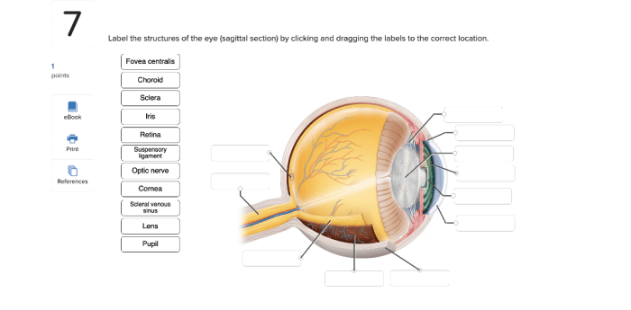
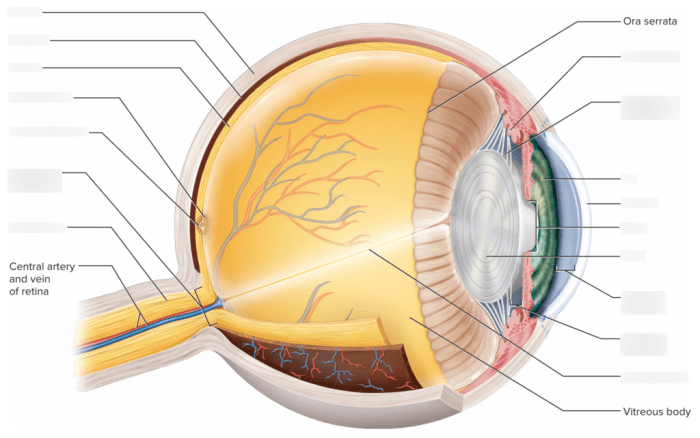
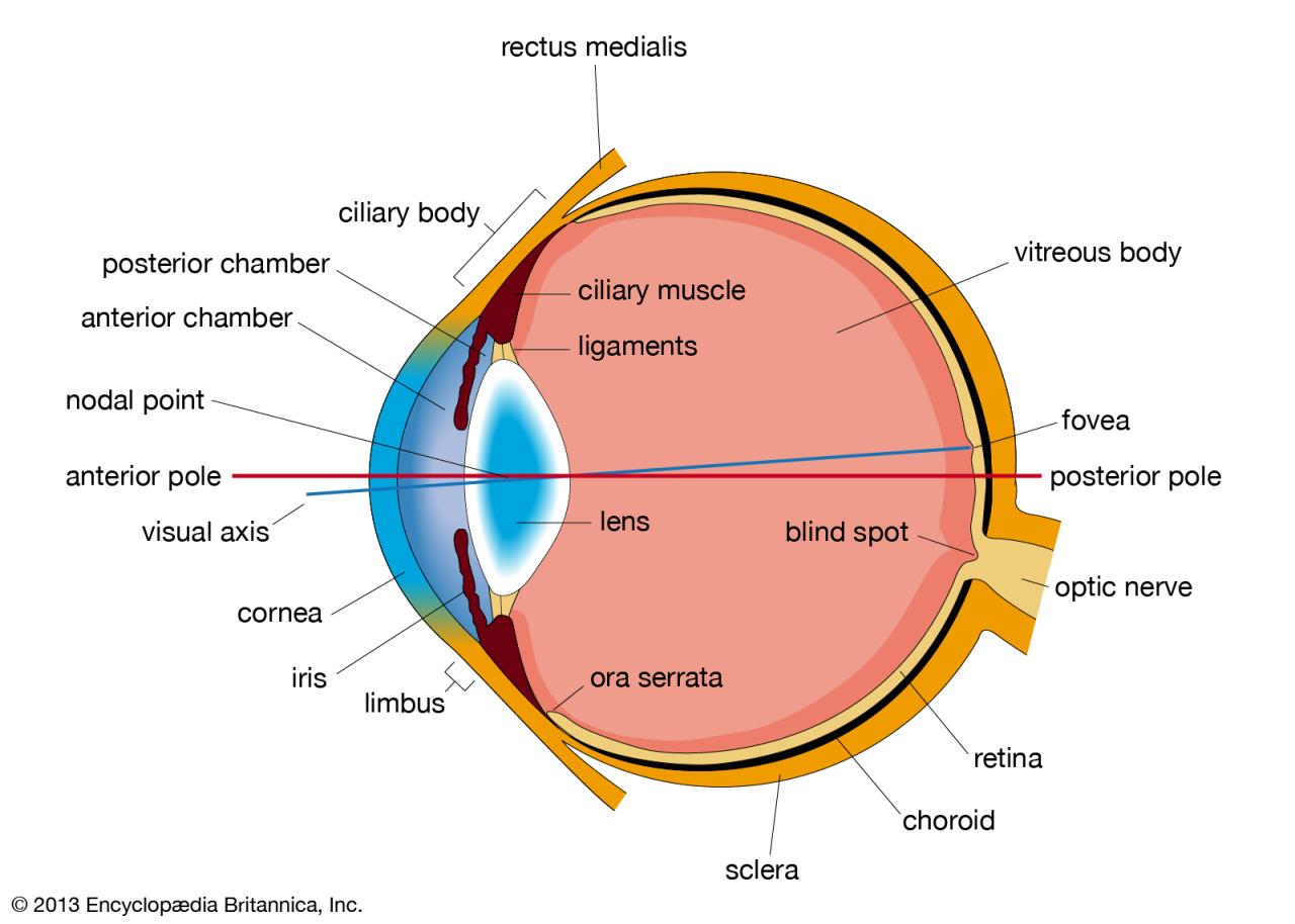
Label the structures in the sagittal section of the eye – Labeling the structures in the sagittal section of the eye provides a comprehensive understanding of the eye’s anatomy. This knowledge is essential for ophthalmologists and optometrists to diagnose and treat various eye conditions.
The sagittal section divides the eye into two symmetrical halves, providing a detailed view of the eye’s internal structures. By examining this section, medical professionals can assess the health of the cornea, lens, retina, and other critical components.
Overview of the Sagittal Section of the Eye

The sagittal section of the eye is a vertical plane that divides the eye into left and right halves. It is an important anatomical landmark used to study the eye’s internal structures and their relationships to each other. The sagittal section is defined by the anatomical planes: the median sagittal plane, which passes through the center of the body, and the coronal plane, which divides the body into anterior and posterior portions.
Labeling the Major Structures: Label The Structures In The Sagittal Section Of The Eye
| Structure | Label | Function | Characteristics |
|---|---|---|---|
| Cornea | A | Transparent, protective covering of the eye | – Avascular
|
| Iris | B | Colored part of the eye that controls pupil size | – Contains sphincter and dilator muscles
|
| Lens | C | Transparent, flexible structure that focuses light on the retina | – Changes shape to accommodate different focal lengths
|
| Pupil | D | Opening in the iris that allows light to enter the eye | – Size controlled by the iris muscles
|
| Retina | E | Light-sensitive layer that converts light into electrical signals | – Contains photoreceptors (rods and cones)
|
| Vitreous Body | F | Gel-like substance that fills the eye’s posterior chamber | – Maintains the shape of the eye
|
| Optic Nerve | G | Transmits visual information from the retina to the brain | – Blind spot (optic disc) where the optic nerve exits the eye
|
| Sclera | H | White, tough outer layer of the eye that protects the inner structures | – Contains collagen fibers
|
Describing the Structures

-
-*Cornea
Transparent, dome-shaped structure that protects the eye and refracts light.
-*Iris
Colored part of the eye that controls pupil size and regulates light entry.
-*Lens
Clear, flexible structure that focuses light on the retina.
-*Pupil
Opening in the iris that allows light to enter the eye.
-*Retina
Light-sensitive layer that converts light into electrical signals.
-*Vitreous Body
Gel-like substance that fills the eye’s posterior chamber and maintains its shape.
-*Optic Nerve
Transmits visual information from the retina to the brain.
-*Sclera
White, tough outer layer that protects the eye’s inner structures.
Histological Features
| Structure | Cellular Composition | Organization | Staining Properties |
|---|---|---|---|
| Cornea | Epithelium, stroma, endothelium | Epithelium: Stratified squamousStroma: Collagen fibersEndothelium: Simple squamous | Epithelium: EosinophilicStroma: BasophilicEndothelium: Acidophilic |
| Lens | Lens fibers | Long, thin, transparent fibers | Basophilic |
| Retina | Photoreceptors, bipolar cells, ganglion cells | Layers: Photoreceptor layer, outer nuclear layer, outer plexiform layer, inner nuclear layer, inner plexiform layer, ganglion cell layer | Photoreceptor layer: Outer segments stain eosinophilicGanglion cell layer: Nuclei stain basophilic |
| Vitreous Body | Collagen fibers, hyaluronic acid | Gel-like network | Eosinophilic |
Clinical Significance

Understanding the sagittal section of the eye is essential for diagnosing and treating various eye conditions:
-
-*Cataracts
Opacities in the lens that can lead to vision loss.
-*Glaucoma
Increased intraocular pressure that damages the optic nerve and can cause blindness.
-*Retinal detachment
Separation of the retina from the underlying choroid, which can lead to vision loss.
-*Macular degeneration
Degeneration of the macula, the central part of the retina responsible for detailed vision.
Top FAQs
What is the clinical significance of understanding the sagittal section of the eye?
Understanding the sagittal section of the eye is crucial for diagnosing and treating various eye conditions. By examining this section, ophthalmologists can assess the health of the cornea, lens, retina, and other structures, helping them identify abnormalities and determine appropriate treatment plans.
What are the key structures visible in the sagittal section of the eye?
The key structures visible in the sagittal section of the eye include the cornea, iris, lens, pupil, retina, vitreous body, optic nerve, and sclera. Each structure plays a specific role in the eye’s function, and abnormalities in any of these structures can indicate underlying eye conditions.
How does the sagittal section of the eye differ from other anatomical sections?
The sagittal section of the eye is unique compared to other anatomical sections because it provides a view of the eye’s internal structures in a symmetrical plane. This section divides the eye into two equal halves, allowing medical professionals to assess the structures in both the anterior and posterior segments of the eye.